

Abstract
Background: Adult T-cell leukemia/lymphoma (ATLL) is a mature T-cell neoplasm, linked to the human T-cell lymphotropic virus, HTLV-1. Patients with ATLL are often at the risk of opportunistic infections. Some studies suggested that ATLL cells originate from HTLV-1-infected regulatory T cells (Tregs). It could be possible that this immunocompromised state is caused by the function of ATLL cells having similar phenotypes with Tregs. In this study, we examined the expression of immunosuppressive molecules associated with Tregs in ATLL cells, and analyzed their roles in the function of ATLL cells.
Methods: The protocol of this study was approved by the Investigational Review Board of Osaka University Hospital. Peripheral blood mononuclear cells (PBMCs) were collected from 10 asymptomatic HTLV-1 carriers and 22 ATLL patients (1 with smoldering type, 5 with chronic type, 2 with lymphoma type, and 14 with acute type) after getting informed consent. PBMCs from 3 ATLL patients were separated into CD4+ CD7- CADM1+ATLL cells and adjacent CD4+CD7+ CADM1-normal T cells using Fluorescence-activated Cell Sorter (FACS), and cells in each fraction were subjected to total RNA sequencing experiments. Based on the results, we examined the expression patterns of CD39 and CD73 in HTLV-1 carriers or each type of ATLL patients, and also analyzed the immune functions of these molecules in ATLL tumor cells.
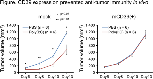
Results: We compared whole transcriptome of ATLL cells and normal CD4+cells. Bioinformatic analyses showed that many genes associated with immunosuppressive functions were elevated or downregulated in ATLL cells. Among these genes we focused on CD39, CD73 and CD26, because they have recently been reported to be strongly associated with the functions of Tregs. CD39, expressed on normal Tregs, and extrinsic CD73 have immunosuppressive potential by catalyzing adenosine from extracellular ATP, and CD26 has opposite potential by resolving adenosine, which have a strong anti-inflammatory function and plays major role in Treg-mediated immunosuppression. We found that all of 4 ATLL cell lines (MJ, MT1, MT2, MT4) expressed CD39, but not CD73 just as human effector Tregs. Tumor cells from 12 acute ATLL patients (86%) and 2 chronic ATLL patients (40%) expressed CD39, but the expressions of CD73 were various. Also in asymptomatic carriers, we could detect CD39 and/or CD73 positive in CD7- CADM1+ abnormal fraction of CD4+cells. On the other hand, CD26, normally expressed on human CD4+Th cells other than effector Tregs, was negative in ATLL cell lines and primary ATLL cells except for cells in abnormal fraction of one asymptomatic carrier. CD39 negative cases in chronic/smoldering type tended to show slower disease progression after the blood collection. Next, the role of CD39 and/or CD73 in ATLL cells was assessed in vitro and in vivo. As expected, CD39+ ATLL cells converted significantly more extracellular ATP than CD39- ATLL cells, and mass spectrometry analysis of AMP/adenosine concentration identified the AMPase activity of CD73+ ATLL cells. Furthermore, we established CD39 knockout (KO) cells from ATL cell-line MJ using CRISPR/Cas9 system, and performed in vitro suppression assays for assessment of immunosuppressive function. Although wild type MJ suppressed the growth of normal CD4+ and CD8+ T cells, KO MJ did little. Next, we analyzed the role of CD39 in the progression of tumor cells in vivo. We transplanted mouse T-cell lymphoma cell-line EG7-OVA artificially expressing CD39 or mock into mice subcutaneously. The coinjection of immunoadjuvant poly(I:C) significantly suppressed the tumor growth of mock cells, but the tumor sizes of CD39 expressing cells were almost the same as those of mock cells without poly(I:C) injection (Figure).
Conclusion: In this study, we reported that most of ATLL cells in acute type patients express CD39+ CD26- just as Tregs, and that CD39- KO of ATLL cell line cancelled its immunosuppressive effects, and forcibly expressed CD39 on tumor cells rejected the anti-tumor immunity in vivo. From these data, we clarified the pathological mechanism of immunosuppressive function in ATLL cells, and also showed that CD39 expression could be used as a prognostic clue and be a new therapeutic target of ATLL.
Ezoe:TAIHO Phamaceutical Co., Ltd.: Research Funding. Yokota:Celgene: Research Funding; Bristol-Myers Squibb: Research Funding; Pfizer Inc.: Research Funding; CHUGAI PHARMACEUTICAL CO., LTD.: Research Funding; MSD K.K.: Research Funding. Ichii:Novartis Pharma K.K.: Speakers Bureau; Kowa Pharmaceutical Co.,LTD.: Speakers Bureau; Celgene K.K.: Speakers Bureau. Shibayama:Novartis Pharma K.K.: Honoraria, Research Funding; Celgene K.K.: Honoraria, Research Funding; Takeda Pharmaceutical Co.,LTD.: Honoraria, Research Funding; Fujimoto Pharmaceutical: Honoraria, Research Funding; Jansen Pharmaceutical K.K: Honoraria; Ono Pharmaceutical Co.,LTD: Honoraria, Research Funding; Mundipharma K.K.: Honoraria, Research Funding; Bristol-Meyer Squibb K.K: Honoraria, Research Funding. Oritani:Novartis Pharma: Speakers Bureau. Kanakura:Alexion Pharmaceuticals, Inc.: Consultancy, Honoraria, Research Funding.

This feature is available to Subscribers Only
Sign In or Create an Account Close Modal